Back Of Skull Anatomy - Sfxkiavdeecw7m : Foramina inside the body of humans and other animals.. The skull includes the upper jaw and the cranium. Looking at it from the inside it can be subdivided into. The skull base is the inferior portion of the neurocranium. The skull also includes cartilage (put your finger on the tip of your nose and wiggle it) and ligaments (open and close your mouth if you want to use them). The frontal, parietal, temporal and occipital bones are joined at the cranial sutures.
The cranium and the mandible. Looking at it from the inside it can be subdivided into. The skull bones can be classified into two groups: Skull reshaping is done on any of the structures that lie above the face. The skull begins to form prior to week 12 of embryogenesis.
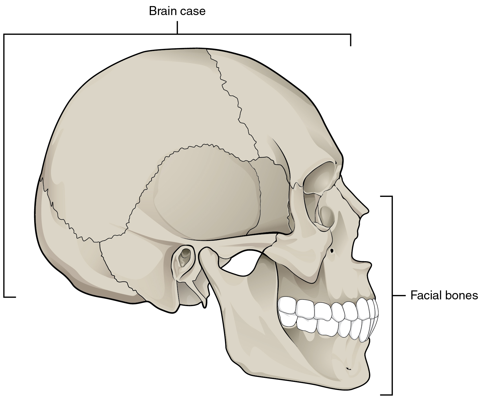
The Skull Anatomy And Physiology from opentextbc.ca Anatomy next provides anatomy learning tools for students and teachers. The skull performs vital functions. Skull reshaping is done on any of the structures that lie above the face. The base of the skull (or skull base) forms the floor of the cranial cavity and separates the brain from the structures of the neck and face. The skull or known as the cranium in the medical world is a bone structure of the head. The cranium and mandible was exported from ct data. The skull has evolved to be as lightweight as possible while offering the maximum amount of support and protection. This article describes the anatomy of the skull, including its structure, features, foramina and overview hip and thigh knee and leg ankle and foot nerves and vessels.
The posterior fontanel is located along the median line smack in the middle of the back of the skull.
The occipital bone forms the back of the skull and the base of the cranium. Learn about the anatomy of the skull bones and sutures as seen on ct images of the brain. It offers protection to the brain, eye balls, inner ears, and nasal passages. Norma basalis ( anterior part , middle part and posterior part ). The skull includes the upper jaw and the cranium. Foramina inside the body of humans and other animals. Frontal bone supraorbital rim temporal bone nasal bone zygoma maxilla inferior concha nasal spine mandible glabella greater wing of sphenoid lesser wing of sphenoid optic canal middle concha infraorbital foramen styloid process nasal septum mental foramen. Cranial cavity , cranial sutures. Skeleton anatomy easy review for practical exam bones and structures. This anatomic region is complex and poses surgical challenges for otolaryngologists and neurosurgeons alike. Anatomical structures of the skull include: The posterior fontanel is located along the median line smack in the middle of the back of the skull. The skull is a bony structure that supports the face and forms a protective cavity for the brain.
It supports and protects the face and the brain. Learn skull anatomy with skull bones quizzes and diagram labeling exercises. Please feel free to download and print. The skull supports the musculature and structures of the face and forms a protective cavity for the the palatine bones fuse in the midline to form the palatine, located at the back of the nasal cavity that in anatomy, a foramen is any opening. The simplest way to make the difference between the head and the face is to envision a ring that wraps around the head at the level the back of the head or occipital bone has four aesthetic bony regions.
Skull Base Imaging I Dre 19 Prof Dr Mamdouh Mahfouz Youtube from i.ytimg.com The skull supports the musculature and structures of the face and forms a protective cavity for the the palatine bones fuse in the midline to form the palatine, located at the back of the nasal cavity that in anatomy, a foramen is any opening. Anatomical structures of the skull include: It was then cleaned, adapted and polypainted this model is part of a comparison with the skull of a human. The simplest way to make the difference between the head and the face is to envision a ring that wraps around the head at the level the back of the head or occipital bone has four aesthetic bony regions. The skull bones can be classified into two groups: This is a model of the human (homo sapiens) skull. Looking at it from the inside it can be subdivided into. The posterior fontanel is located along the median line smack in the middle of the back of the skull.
The major sutures are the coronal suture, sagittal suture, lambdoid suture and squamosal sutures.
The occipital bone forms the back of the skull and the base of the cranium. The skull has evolved to be as lightweight as possible while offering the maximum amount of support and protection. Overview, anterior skull base, middle skull base march 18, 2017. The skull is the bony skeleton of the head. Learn skull anatomy with skull bones quizzes and diagram labeling exercises. It is comprised of many bones, formed by intramembranous ossification, which are joined together by sutures (fibrous joints). Human skull from the front. Some bones give shape to the face, others protect the brain. The skull begins to form prior to week 12 of embryogenesis. The major sutures are the coronal suture, sagittal suture, lambdoid suture and squamosal sutures. The bbc is not responsible for the content of external websites. It was then cleaned, adapted and polypainted this model is part of a comparison with the skull of a human. The two fontanels located on the sides of the skull are mirror.
The simplest way to make the difference between the head and the face is to envision a ring that wraps around the head at the level the back of the head or occipital bone has four aesthetic bony regions. The skull begins to form prior to week 12 of embryogenesis. Anatomy of the skull and bones of cranium on medical illustrations. Excluding ear ossicles, it is made of 22 bones. Skeleton anatomy easy review for practical exam bones and structures.
Skull Base Imaging I Dre 19 Prof Dr Mamdouh Mahfouz Youtube from i.ytimg.com This is a model of the human (homo sapiens) skull. Learn vocabulary, terms and more with flashcards, games and other study tools. In order to be light, the skull is made up by flat and irregular bones, and has hollow spaces called the sinuses. The skull is the bony skeleton of the head. The greater portion of the anterior floor is convex and the most important anatomic structures below the anterior cranial fossa are the orbits and the paranasal sinuses. So, the human skull consists of 23 bones. Looking at it from the inside it can be subdivided into. Learn about the anatomy of the skull bones and sutures as seen on ct images of the brain.
Norma basalis ( anterior part , middle part and posterior part ).
Overview, anterior skull base, middle skull base march 18, 2017. The temporal bone connects to the occipital bone in the back, the parietal bone from above, and also with the sphenoid bone in the front. Excluding ear ossicles, it is made of 22 bones. It supports and protects the face and the brain. The skull has a single occipital condyle.7 the skull consists of five major bones: The skull begins to form prior to week 12 of embryogenesis. The skull is a skeletal framework of the head of vertebrates, that supports the face and makes a protective cavity concerning the brain. The bone is pierced by a large oval hole(the foramen magnum) through which runs the spinal cord. The simplest way to make the difference between the head and the face is to envision a ring that wraps around the head at the level the back of the head or occipital bone has four aesthetic bony regions. Learn vocabulary, terms and more with flashcards, games and other study tools. This anatomic region is complex and poses surgical challenges for otolaryngologists and neurosurgeons alike. Anatomical structures of the skull include: But it's not all bones!


0 Komentar